
Fig. 3. Testing whole blood.
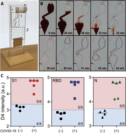
(A) Modified microfluidic flow cell for testing whole blood. Zone 1: The reaction chamber was modified to prevent red blood cells from collecting in the chamber. Zone 2: The incubation timing channel was shortened to compensate for the slower flow rate of blood and to ensure blood did not clot or clog the channels. (B) Time lapse of blood and wash buffer in the reaction chamber. (C) Aggregated data for five positive samples and four negative controls tested for antibodies against S1, RBD, and N. Dotted lines represent 2 SDs above the mean of the negative controls; 100% sensitivity (5/5) and 100% specificity (4/4) were achieved for S1, RBD, and N. Photo credit for (A) and (B): David S. Kinnamon, Duke University.