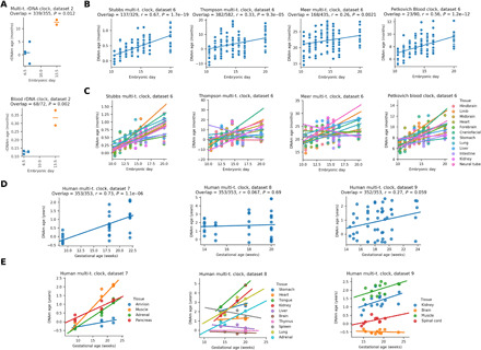
Fig. 2. Organismal aging begins in mid-embryonic development in mice and humans.

(A) Epigenetic age (multi-tissue and blood rDNA clocks) analysis of the dataset that contains both early and late mouse embryo samples (E13.5 samples are based on primordial germline cells). (B) Application of genome-wide epigenetic clocks to later stages of mouse embryogenesis (r, Pearson correlation coefficient; P, P value of the correlation). (C) The same data as above but separated by tissue. An increasing trend is observed for almost all tissues, with few nonsignificant exceptions. (D) Epigenetic age dynamics of four independent prenatal human 450K methylation array datasets based on the Horvath human multi-tissue clock. (E) The same data as above but separated by tissue (five significant increases, nine nonsignificant increases, four nonsignificant decreases, and zero significant decreases).
