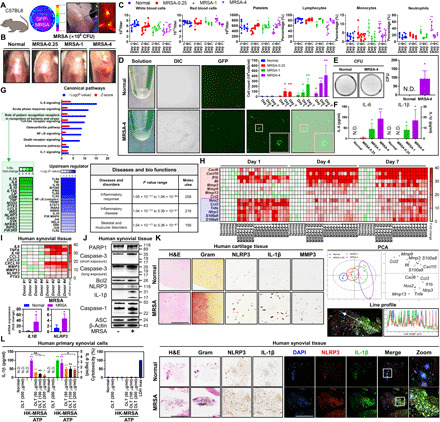
Fig. 1. MRSA-induced septic arthritis results in inflammation, cartilage degradation, and NLRP3 inflammasome activation in human and murine knee joint tissue.

(A) C57BL/6 mice were intra-articularly injected with Dulbecco’s phosphate-buffered saline (DPBS), GFP-MRSA 0.25 × 10⁶ colony-forming units (CFU) (MRSA-0.25), GFP-MRSA 1 × 10⁶ CFU (MRSA-1), and GFP-MRSA 4 × 10⁶ CFU (MRSA-4) (n = 3 to 6 per group). (B) Representative knee images at 7 days. (C) Blood was collected at 1, 4, and 7 days for complete blood count (CBC) quantification. (D) MRSA were detected in synovial fluid and through differential interference contrast (DIC) as well as GFP labeling and (E) MRSA growth was quantified. (F) Synovial IL-6 and IL-1β expression was measured. (G) Whole-genome transcriptomic profiling of MRSA-infected bone marrow–derived macrophages was performed. Upstream regulator is indicative of the transcriptional regulators driving the observed gene expression changes. (H) Synovial mRNA expression was analyzed by principal components analysis (PCA) (fig. S1A). (I to L) Human synovium and cartilage were incubated with MRSA-4 for 24 hours (n = 4 each). (I) mRNA expression was examined (fig. S1B). (J) PARP1, caspase-3, Bcl2, NLRP3, IL-1β, caspase-1, ASC, and β-actin expression was quantified using β-actin as a control. (K) Tissue sections were visualized with hematoxylin and eosin (H&E) and Gram stains, and NLRP3, IL-1β, and MMP3 expression was detected. Colocalization of NLRP3 and IL-1β was confirmed by line profile analysis (fig. S2). (L) IL-1β, IL-6, and cytotoxicity levels in HK-MRSA–primed, ATP-stimulated synovial cells after OLT treatment. Error bars represent means ± SD. One-way analysis of variance (ANOVA) with Tukey’s post hoc analysis was performed (*P < 0.05 or **P < 0.01; N.D., not detected; scale bar, 1000 μm). DAPI, 4′,6-diamidino-2-phenylindole. Photo credit: Hyuk-Kwon Kwon, Yale University.
