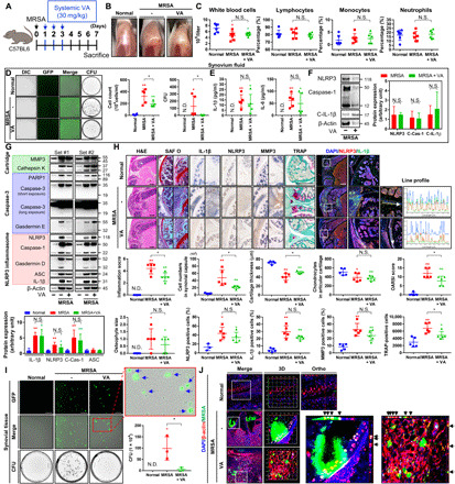
Fig. 2. Treatment with vancomycin is effective for septic arthritis caused by MRSA, but MRSA persists intracellularly and induces a proinflammatory state.

(A) C57BL/6 mice were intra-articularly injected with DPBS or MRSA (4 × 10⁶ CFU). One day after infection, vancomycin (VA; 30 mg/kg) was subcutaneously injected for 3 days (n = 3 to 5 per group). (B) Representative images of knees. (C) Blood was collected, and CBCs were obtained. (D) Synovial cells and GFP-labeled MRSA CFU were quantified. (E) Synovial IL-1β and IL-6 concentrations were quantified with or without systemic vancomycin. (F) Synovial fluid NLRP3, caspase-1, C-IL-1β, and β-actin expression was measured using β-actin as a loading control. (G) Synovial MMP3, cathepsin K, PARP1, caspase-3, GSDME, NLRP3, caspase-1, GSDMD, ASC, IL-1β, and β-actin expression was measured with a β-actin control. (H) Tissue sections were stained with H&E, safranin O (SAF O), and TRAP. The percentage of NLRP3-, IL-1β–, and MMP3-positive cells were quantified, and TRAP-positive cells were counted (scale bar, 1000 μm) (fig. S3A). Coexpression and colocalization of NLRP3 and IL-1β were confirmed by line profile analysis (scale bar, 1000 μm) (fig. S3B). (I) MRSA CFU quantification from single synovial tissue cells. The blue arrow indicates intracellular MRSA (scale bar, 100 μm). (J) Intracellular and extracellular MRSA were measured by multiplex immunohistochemistry and confirmed using three-dimensional (3D) and orthogonal analyses (fig. S4). Error bars show means ± SD. One- or two-way ANOVA with Tukey’s post hoc analysis was used (*P < 0.05 or **P < 0.01; N.S., not significant). Photo credit: Photographer name: Hyuk-Kwon Kwon. Photographer institution: Yale University.
