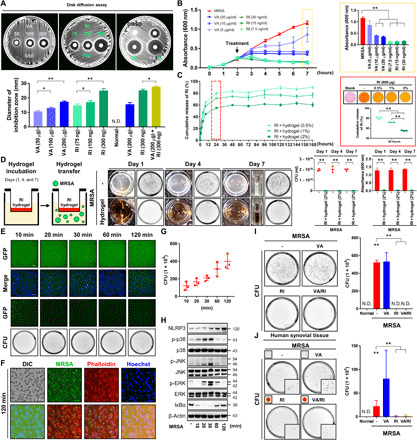
Fig. 4. Rifampin-containing hydrogels are effective in removing intracellular MRSA.

(A) Disk fusion assays using vancomycin (20 to 200 μg), rifampin (RI; 75 to 300 ng), and vancomycin (200 μg) and rifampin (300 ng). Inhibition zone diameters were measured. DMSO, dimethyl sulfoxide. (B) MRSA (4 × 10⁶ CFU) was seeded and grown for 2 hours and then treated with vancomycin (5 to 20 μg/ml) and rifampin (7.5 to 30 ng/ml). Absorbance levels are shown. (C) The release efficiency of rifampin (600 μg) from hydrogel (0.5 to 2%) was measured as cumulative rifampin release over time. (D) Two percent rifampin (600 μg) hydrogel was added in polyester (PET) track-etched membrane inserts and then transferred to lysogeny broth media containing 4 × 10⁶ CFU MRSA after 1, 4, and 7 days. MRSA growth was measured via absorbance and CFU. (E) MRSA (4 × 10⁶ CFU) was cocultured with RAW264.7 cells for 10 to 120 min, after which intracellular fluorescence was measured (fig. S7A) (scale bar, 100 μm). (F) Cells were stained with Phalloidin and Hoechst 120 min after infection (scale bar, 27 μm). (G) MRSA bioburden in this cellular suspension was counted (fig. S7B). (H) NLRP3, p38, c-Jun N-terminal kinase (JNK), extracellular signal–regulated kinase (ERK), inhibitor of nuclear factor κBα (IκBα), and β-actin expression was measured using β-actin as a control. (I) MRSA-infected cells were treated with vancomycin (300 μg/ml) and/or rifampin (60 μg/ml) 120 min after infection. Intracellular MRSA was quantified. (J) Human synovium was infected with MRSA (4 × 10⁶ CFU) for 4 hours and then treated with vancomycin-loaded (300 μg/ml) and/or rifampin-loaded (60 μg/ml) hydrogel. Intracellular MRSA was quantified. Error bars show means ± SD. One- or two-way ANOVA with Tukey’s post hoc analysis was performed (*P < 0.05 and **P < 0.01). Photo credit: Photographer name: Hyuk-Kwon Kwon. Photographer institution: Yale University.
