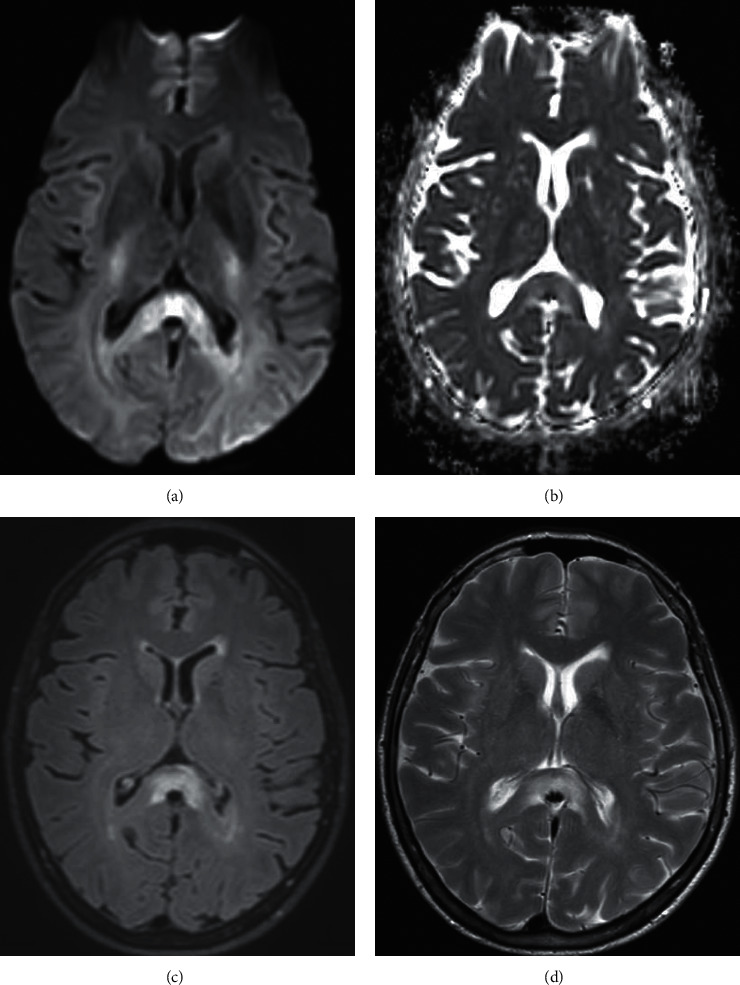
Figure 2.

MR brain with and without contrast at one month follow up. (a) DWI, axial cut. (b) ADC axial cut. (c) FLAIR axial cut. (d) T2 axial cut. In comparison, the corpus callosum lesion is stable compared to prior imaging with the absence of novel interval lesions.
