Fig. 1.
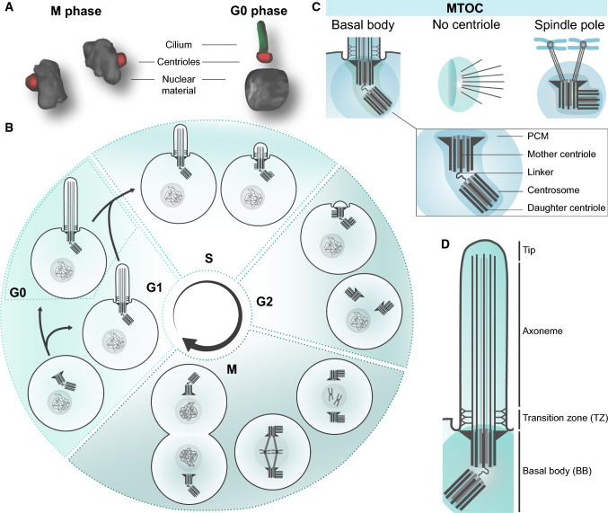
Cilia and ciliation cycle. a Graphic representation of the centrioles. During mitosis (M phase) the centrioles form the spindle poles to separate the nuclear material. In quiescence (G0 phase) the centrioles (red) are positioned at the base of the cilium (green). b The cilia assemble every cell cycle in G0/G1 phase and disassemble during S/G2 phase, during which the centrioles are positioned at the ciliary base. After detachment from the plasma membrane, the linker between the mother and daughter centriole dissolves, allowing the two centrioles to move towards the nucleus to form the spindle poles. Each mother centriole forms a new daughter centriole during the next cycle. c The centrosomes, displaying two structurally different centrioles surrounded by the pericentriolar material (PCM), act as the main MTOC both during spindle pole formation and ciliogenesis. Nonetheless, an MTOC can also arise without a centrosome [174]. The spindle pole and basal body refer to the centrosome as an MTOC with the pericentriolar material, but either contain proteins specific to the cell cycle, or ciliogenesis. The centrosomes gather different complexes for these specific functions, enriching the PCM for different sets of proteins, and making it more likely for interacting proteins to bind at the right phase of the cell cycle. d Schematic representation of the cilia and the ciliary regions that can be distinguished

