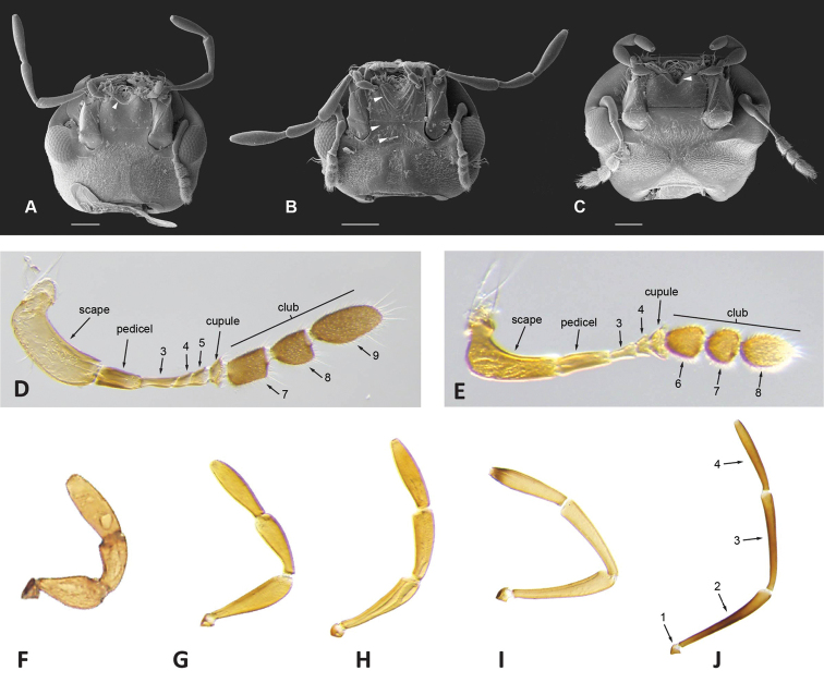
Figure 12.
Head structures A–C scanning electron micrographs of ventral view of head: ATobochares pallidus with smooth mentum and white arrow pointing to transverse carina limiting posterior margin of antero-medial depression BNanosaphes tricolor with top white arrow pointing to oblique crenulations of mentum, mid white arrow pointing to flat and smooth anterior surface of submentum, and bottom white arrow pointing to concave posterior surface of submentum CQuadriops reticulatus with white arrow pointing to antero-medial depression of mentum D, E light micrographs of antenna: DAulonochares tubulus (9 antennomeres) EChasmogenus cremnobates (8 antennomeres) F–J light micrographs of maxillary palps: FQuadriops reticulatusGAgraphydrus insidiatorHHelochares sp. IHelochares lividusJAulonochares tubulus. Scale bars: 100 μm (A–C)

