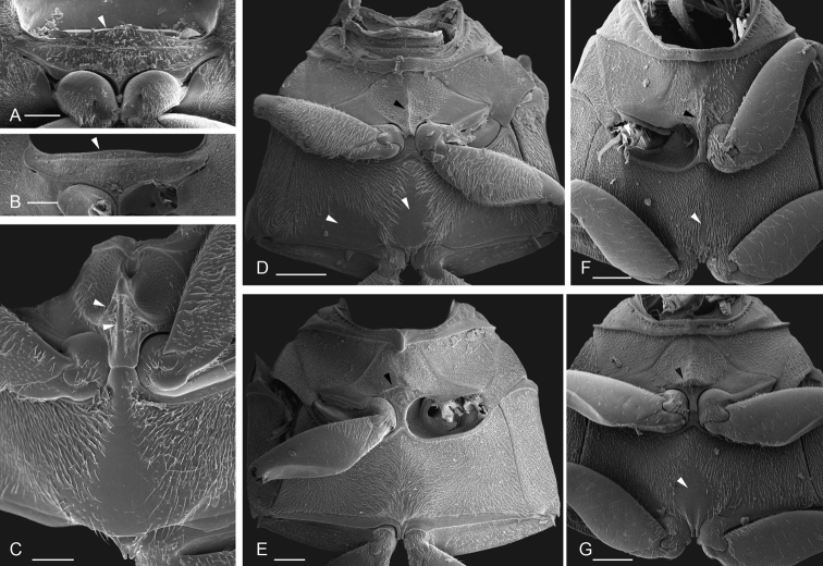
Figure 14.
Scanning electron micrographs of thorax in ventral view A, B prosternum: ATobochares striatus with white arrow pointing to anterior projection BQuadriops reticulatus with white arrow pointing to anterior projection C–G mesoventrite and metaventrite: CCrucisternum ouboteri with white arrows pointing to anteriorly pointed transverse ridge and longitudinal carina of mesoventrite, metaventrite with median glabrous patch DNanosaphes tricolor with black arrow pointing to longitudinal carina along mesoventrite and white arrows pointing to median and postero-lateral glabrous patches of metaventrite EQuadriops reticulatus with black arrow pointing to transverse carina across mesoventrite and metaventrite uniformly pubescent FTobochares communis with black arrow pointing to longitudinal carina along mesoventrite and white arrow pointing to narrow postero-medial glabrous patch on metaventrite GTobochares kasikasima with black arrow pointing to transverse elevation across mesoventrite and white arrow pointing to broad postero-medial glabrous patch on metaventrite. Scale bars: 100 μm.

