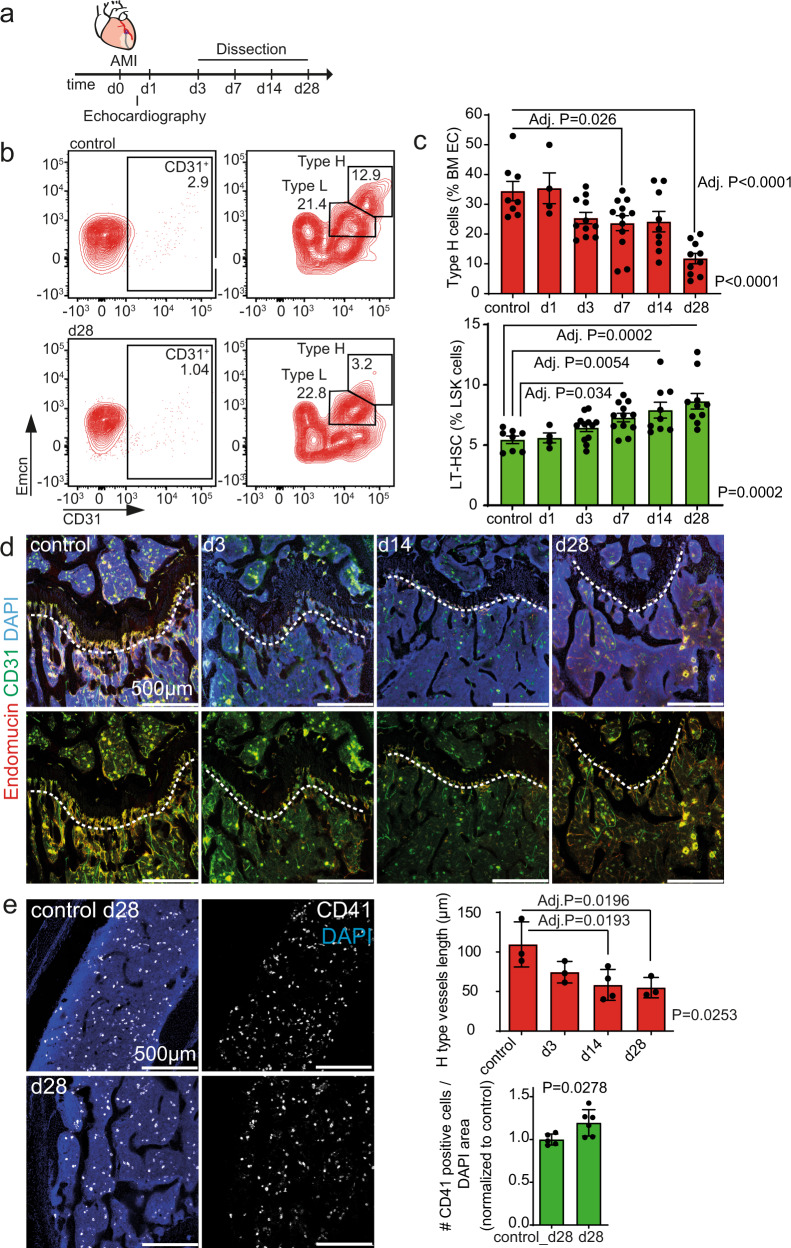
Fig. 1. Bone type H endothelium is reduced upon myocardial infarction (MI).
a Schematic of the experimental design. b, c Flow cytometry analysis of femur bone marrow. Surgery was performed on 12-week-old animals. b Gating strategy for endothelial cells (CD45negTer119neg viable single bone cells are shown with further gating on CD31pos cells, followed by gating of Type H and Type L EC subsets, based on CD31 and Emcn expression). The numbers shown within the gates represent the percentages of events (cells) within that gates relative to the upstream parent gates (CD45negTer119neg for CD31pos cells and CD31pos cells for Type H and Type L EC subsets, accordingly). Representative samples from control and day (d) 28 are shown. c Quantification. Upper panel, type H endothelial cells are reduced relative to all bone marrow endothelial cells (BM EC) after MI. Lower panel, Long-term hematopoietic stem cells (LT-HSC) are increased relative to LSK cells after MI. N = 8 for control, N = 4 for d1, N = 12 for d3, N = 12 for d7, N = 9 for d14, and N = 10 for d28. Data are shown as mean ± SEM. P-value was calculated with ANOVA with Dunnet’s multiple comparison test. d, e Immunostaining of longitudinal sections through the femur. d The length of type H vessels (measured in µm, indicated by the dashed line) is reduced after MI. N = 3 for control, d3 and d28, and N = 4 for d14. Data are shown as mean ± SEM. P-value was calculated with ANOVA. Comparisons among all groups were calculated by Dunnet’s multiple comparison test. e Myeloid progenitor cell number is increased in the bone marrow 28 days after MI. N = 5 for each condition. Data are shown as mean ± SEM. P-value was calculated by unpaired, two-tailed Student’s t-test.

