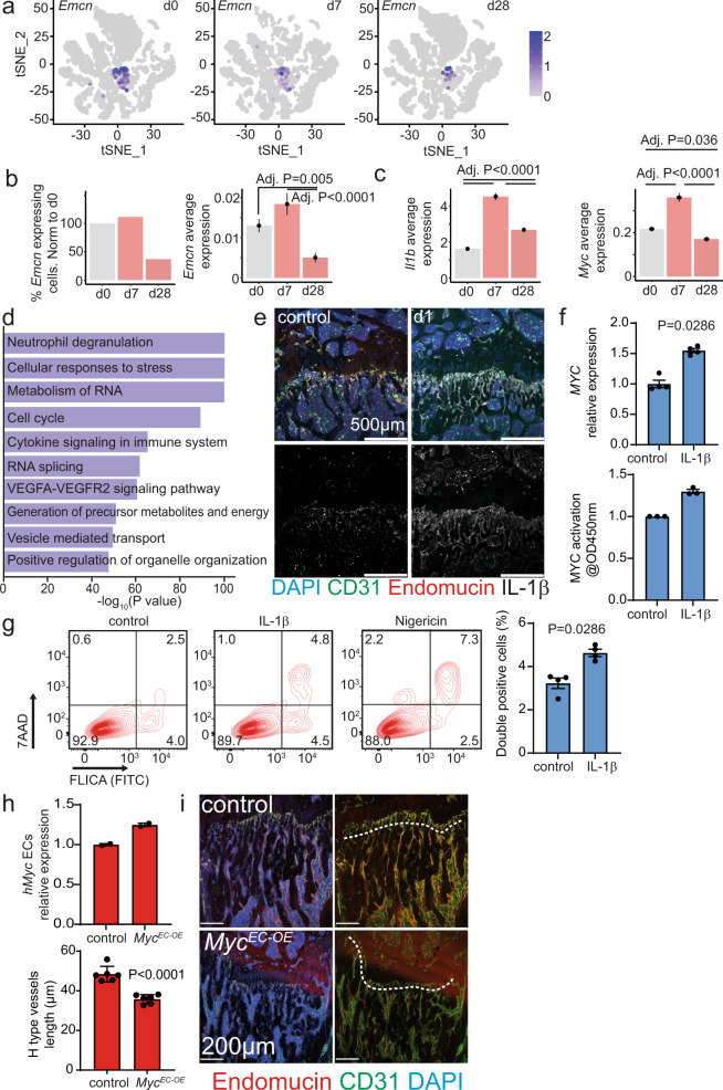
Fig. 3. IL-1β induces MYC and pyroptosis in endothelial cells.
a scRNA-seq of mice bone marrow vascular niche 7 (d7) and 28 days (d28) after myocardial infarction as well as of controls (d0). Clustered cells from the three-time points are displayed in t-SNE plots. Emcn expressing cells are colored. b-left Representation of the percentage of Emcn expressing cells of the lineage depleted, CD31 expressing population. b-right Emcn and (c) Il1b and Myc average expression. d Representation of the ten most significant upregulated terms at d7 revealed by gene ontology analysis when comparing d0 to d7. e Immunostaining of longitudinal femur sections at d1. IL-1β immuno-signal is enriched in type H vessels one day after the ischemic insult. f-up RT-qPCR analysis of MYC expression in HUVEC cells after 1 h IL-1β treatment. N = 4. Data are shown as mean ± SEM. P-value was calculated by a two-tailed Mann–Whitney test. f-down Analysis of MYC activation in HUVEC cells after 1 h IL-1β treatment. N = 3 independent experiments with two technical replicates. Data are shown as fold-change relative to control. g Analysis of Caspase1 activation in HUVECs after 1 h IL-1β treatment or nigericin as a positive control. Left, Gating strategy. Right, quantification. N = 4. Data are shown as mean ± SEM. P-value was calculated by a two-tailed Mann–Whitney test. h Analysis of human MYC expression in isolated liver endothelial cells by RT-qPCR. N = 2 for both groups. Data are shown as mean ± SEM. i Immunostaining of longitudinal sections through the femur. The length of type H vessels (indicated by the dashed line, endomucin in red, CD31 in green, and DAPI in blue) is reduced in MycEC-OE mice when compared to controls. Tamoxifen was given at 8 weeks and analysis performed 4 weeks later, N = 6 for both groups. Data are shown as mean ± SEM. P-value was calculated by unpaired, two-tailed Student’s t-test.

