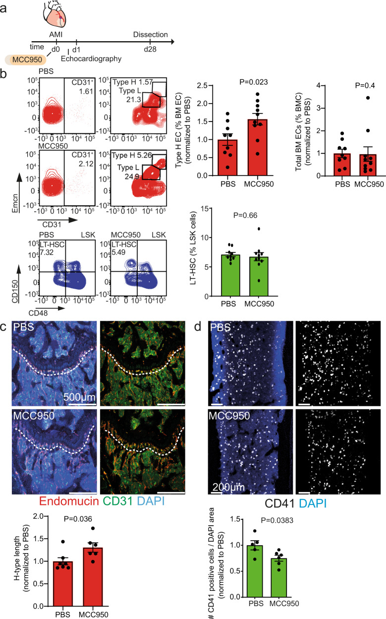
Fig. 4. Anti-IL-1β treatment ameliorates the loss of type H vasculature in the bone after MI.
a Schematic of the experimental design. b Flow cytometry analysis of femur bone marrow. Left, gating strategy; right, quantification. The numbers shown within the gates represent the percentages of events (cells) within that gates relative to the corresponding parent gate. The numbers shown within the gates represent the percentages of events (cells) within that gates relative to the upstream parent gates (CD45negTer119neg for CD31pos cells and CD31pos cells for Type H and Type L EC subsets, accordingly) (red). Type H endothelial cell number is increased upon anti-IL-1β treatment (MCC950), while the total number of endothelial cells is not affected. N = 9 for PBS and N = 10 for MCC950. Data are shown as mean ± SEM, normalized to PBS. P-value was calculated by unpaired, two-tailed Student’s t-test (type H endothelial cell number) and two-tailed Mann–Whitney test (total number of endothelial cells). EC,# endothelial cells, BM EC, bone marrow endothelial cells (green). The total number of long-term hematopoietic stem cells (LT-HSC) remains unchanged after IL-1β treatment. N = 9 PBS and N = 10 MCC950. Data are shown as mean ± SEM. P-value was calculated by unpaired, two-tailed Student’s t-test. LSK, Lin–Sca-1+c-kit+ cells (c) and (d) Immunostaining of longitudinal femur sections. c The length of type H vessels (dashed line) is longer upon MCC950 treatment 28 days after MI. N = 7 for PBS and N = 6 for MCC950. Data are shown as mean ± SEM. P-value was calculated by unpaired, two-tailed Student’s t-test. d CD41+ myeloid progenitor cell number is decreased in the bone marrow of MCC950-treated animals after myocardial infarction. N = 5 PBS and N = 6 MCC950. Data are shown as mean ± SEM. P-value was calculated by unpaired, two-tailed Student’s t-test.

