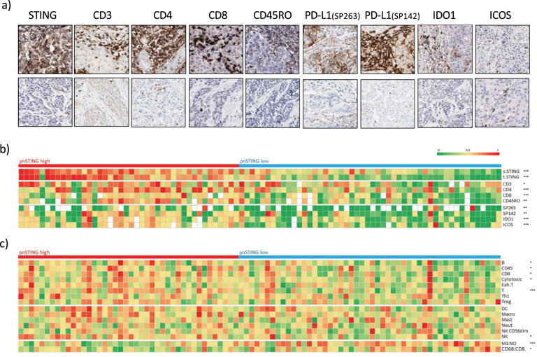
Fig. 3. pnSTING and immune correlates in ER + breast cancer.
a Representative IHC images showing high (top panel) and low (lower panel) expression of perinuclear STING, CD3, CD4, CD8, CD45RO, PD-L1 (SP263), PD-L1 (SP142), IDO1 and ICOS. b Heatmap of normalized expression measured by IHC of perinuclear STING, stromal STING, tumor STING, CD3, CD4, CD8, CD45RO, PD-L1 measured by SP263 and SP142, IDO1 and ICOS in ER + breast cancer cases. c Heatmap of normalized immune scores derived from deconvolution of microarray data in ER+ breast cancer cases. Correlation between markers and pnSTING stratified based on high (above median) and low (below median) was assessed using the Krushall Wallis test on non-transformed data with *, **, and *** indicating a p values of <0.05, <0.01, and <0.001, respectively.

