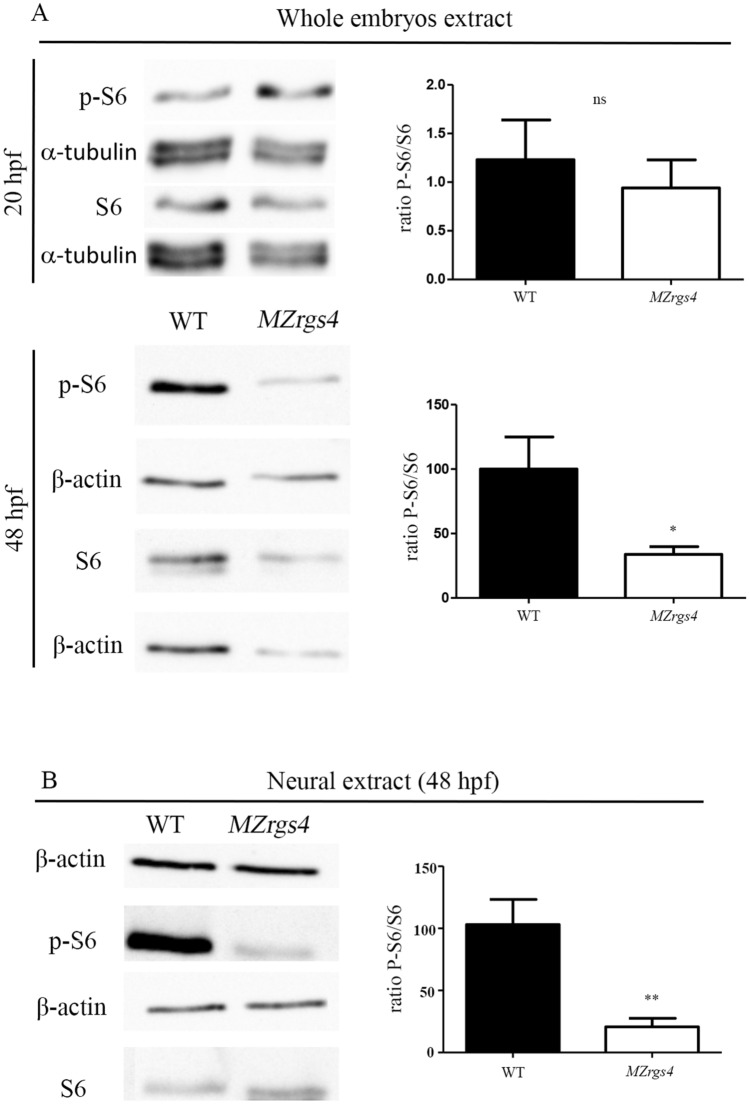
Figure 7.
Loss of rgs4 function alters mTOR signaling in neural cells (A) Immunoblotting of lysates from zebrafish embryos at 20 hpf. S6 and p-S6 amounts were normalized to α-tubulin and the ratio of p-S6 relative to S6 was compared between WT and MZrgs4 lysates. No significant difference was observed between the two groups. Immunoblotting of lysates from zebrafish embryos at 48 hpf. S6 and p-S6 amounts were normalized to β-actin and the ratio of p-S6 relative to S6 was compared between WT and MZrgs4 lysates. MZrgs4 embryos show a significant decrease in the amount of p-S6/S6 in comparison to WT. (B) Immunoblotting of neural lysates from zebrafish embryos at 48 hpf. S6 and p-S6 amounts were normalized to β-actin and the ratio of p-S6 relative to S6 was compared between WT and MZrgs4 lysates. MZrgs4 embryos showed a significant decrease in the amount of p-S6/S6 in comparison to WT.

