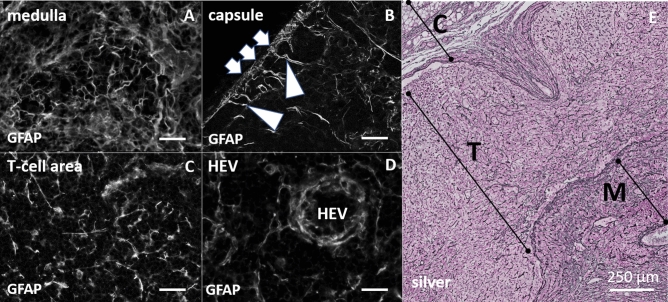
Figure 2.
Left site (A–D), immunohistochemical staining with polyclonal anti-GFAP antibody (DAKO) in different areas of the lymph node, colorless display for impartial interpretation. (A) Undirected arrangement of branched structures in the medulla. (B) Clear signals in the capsule (arrow). Radially symmetric inward extending of longer directional signals (arrowheads). (C) Heterogeneous signal distribution with clearly reticular arrangement of signals in the T-cell area. (D) Concentric and dense packed signals around the high endothelial venules (HEV) (Scale bar for A–D 50 µm). Right site (E) silver staining of lymph node cross section. (E) Digital enlargement of a silver-stained cross-section through a lymph node. Magnification covers as much as possible areas from the capsule (C), T-cell region (T) and the medulla (M). The fibrous structures of this staining represent the reticular network in the lymph node.

