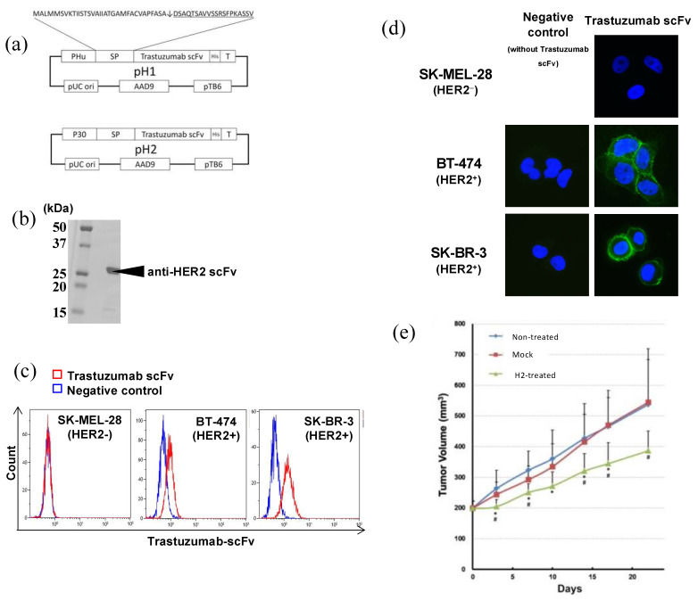
Figure 5.
(a) Structure of the pH1 and pH2 plasmids. (b) Molecular size of the trastuzumab scFV produced by Bifidobacterium. Results for H1 scFv are shown. Regarding H2, identically sized scFv was confirmed at a markedly higher expression amount. (c) FACS analysis. SK-MEL-28 (HER2−), BT-474 (HER2 +/−), and SK-BR-3 (HER2+) cells were stained with His-tag-purified trastuzumab scFv. Blue line: control (buffer alone). Red line: stained with trastuzumab scFv from H2. (d) Immunostaining of cultured cells by His-tag-purified trastuzumab scFv from B. longum H2. Immunofluorescent staining. Blue: nucleus. Green: stained with trastuzumab scFv from H2. Right panels: stained with trastuzumab scFv. Left panels: negative control (without trastuzumab scFv). Original magnification of all images was x400. (e) Growth suppression of a human HER2(+) carcinoma transplanted into nude mice by recombinant Bifidobacterium H2. B. longum mock and H2 were i.v. administered to NCI-N87 human gastric cancer tumor-bearing mice twice a week. Mean ± standard deviation values of 8 mice. *: p < 0.05 versus non-treated group, #: p < 0.05 versus mock-treated group. This figure was adopted from a previous study [58].

