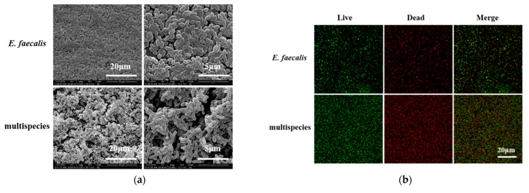
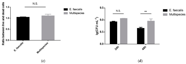
Figure 4.
The biofilm formation of E. faecalis and multispecies models. (a) The scanning electron microscopy images of E. faecalis and multispecies biofilms with 5000× and 20,000× magnified visual fields; (b) the live/dead bacterial staining image of biofilms (live bacteria, stained green; dead cells, stained red); (c) the ratio of the live bacteria cells to dead cells was computed in line with 3 random sights of biofilms; (d) colony-forming units of bacteria in biofilms formed by E. faecalis and multispecies (n = 3). Data are presented as the mean ± standard deviation (** p < 0.01).


