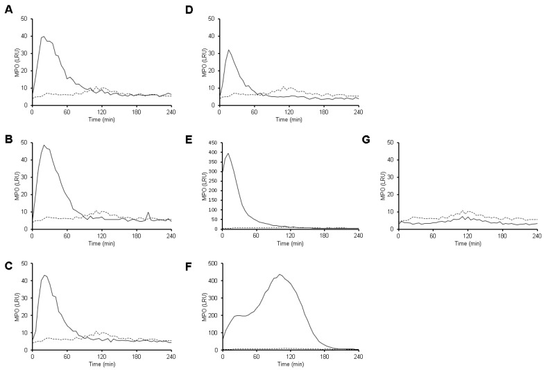
Figure 6.
MPO activity is detected during interaction between neutrophils with E. histolytica. Neutrophils (1 × 10⁵) were culture in RPMI-1640 medium supplemented with 5%FBS and luminol (200 µM). Cells were stimulated with viable E. histolytica trophozoites at ratios of 1:100 (A), 1:50 (B), 1:20 (C) and 1:10 (D), as well as 50 nM PMA (E), 10 µM A23187 (F). Amoebas alone (1 × 10⁴) also were tested (G). Black line represents MPO activity (as luminescence relative units, LRU) after stimulation and dotted line represents MPO activity on neutrophils in the absence of stimuli (same for all plots). Values are means of three independent experiments.

