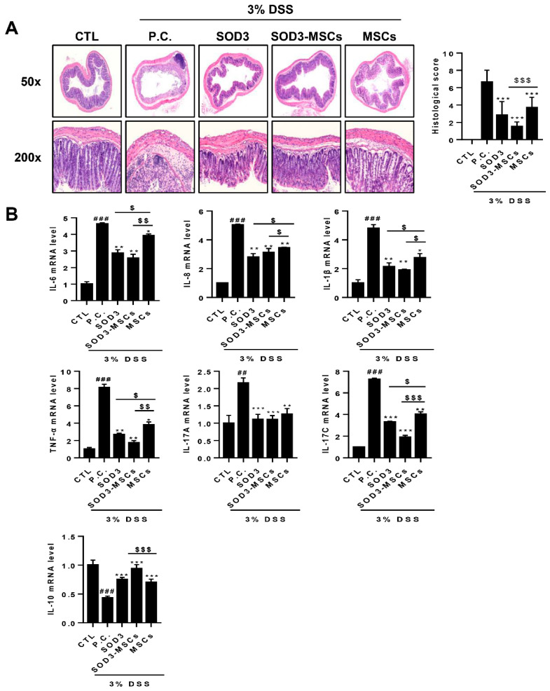
Figure 4.
Histological and molecular analysis of colon samples from SOD3- or SOD3-MSC-treated mice. Colon sections were prepared and H&E staining was performed. (A) H&E stained sections were photographed and representative pictures are shown. Histopathological scores were measured by analyzing epithelial destruction and lymphocyte infiltration. (B) The expressions of pro- or anti-inflammatory genes in the colon were determined by real-time PCR. ## p < 0.01, ### p < 0.001 (CTL vs. P.C), * p < 0.05, ** p < 0.01, *** p < 0.001 (P.C vs. SOD3, SOD3-MSCs and MSCs), $ p < 0.05, $$ p < 0.01, $$$ p < 0.001 (SOD3, SOD3-MSCs vs. MSCs).

