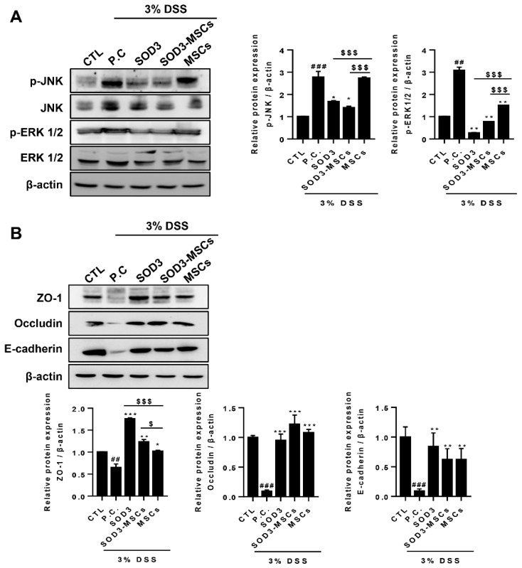
Figure 5.
Expression of proteins in MAPK pathway and epithelial junctions in colons of colitis mice. (A) p-JNK, JNK, p-ERK1/2, and ERK1/2 were detected by immunoblotting. The expressions of p-JNK and p-ERK1/2 were quantified. (B) Expressions of ZO-1, occludin, and E-cadherin were analyzed by immunoblotting and quantified. ## p < 0.01, ### p < 0.001 (CTL vs. P.C), * p < 0.05, ** p < 0.01, *** p < 0.001 (P.C vs. SOD3, SOD3-MSCs and MSCs), $ p < 0.05, $$$ p < 0.001 (SOD3, SOD3-MSCs vs. MSCs). Results show one representative experiment of at least three independent experiments.

