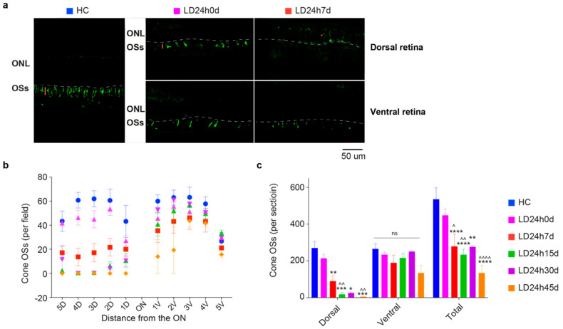
Figure 8.
Light-induced damage to the L/M cone-photoreceptors population. (a) Retinal cross-sections from a representative HC, and 0 and 7 days after light-induced retinal damage. Sections are labeled with an antibody against L/M opsin. Outer segments (OSs) appear normally distributed throughout the retina in the control condition. Light-exposed retinas immediately showed a partial reduction of cone density (0 d) depicted by the length of the orange bar and redistribution of the signal into the soma (*, orange) in the dorsal retina, shortening the remaining OSs in the ventral at 7d. Abbreviations: ONL: outer nuclear layer; OSs: outer segments. Scale bar = 50 µm. (b,c) Numbers of cones distributed along the retinal sections passing through the optic nerve (ON), the totals on the dorsal and ventral sides, and their sum for HC, LD24h0d, LD24h7d, LD24h15d, LD24h30d, and LD24h45d. Statistical significance is represented as follows: *,^ p ≤ 0.05, **,^^ p ≤ 0.01, *** p ≤ 0.001, ****,^^^^ p ≤ 0.0001; * and ^ refer to HC and LD24h0d, respectively; two-way ANOVA with Tukey’s post hoc test. Data represent the means ± SEM of at least N = 5 retinas from different rats, for each experimental condition.

