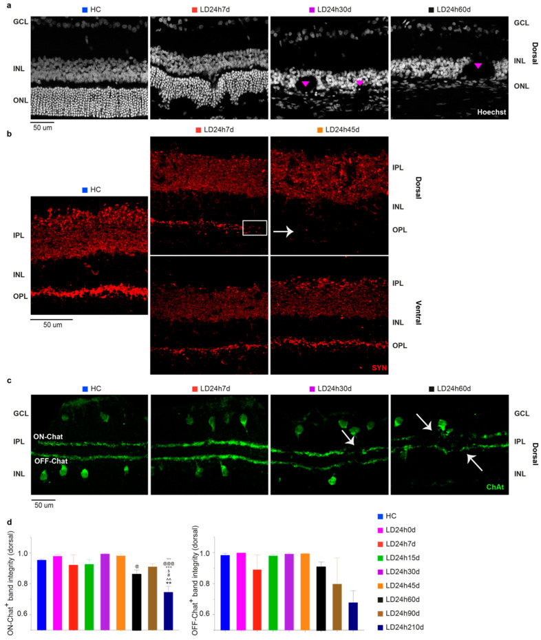
Figure 9.
Inner retinal changes at different recovery times following 24 h of light exposure. (a) Hoechst-stained retinal cross-sections from the dorsal (hot spot) retina. A considerable disruption of the integrity of the INL is depicted. Structures like rosettes were formed (pink triangles) (see Figure 7b; INL holes were not present in the ventral retina). (b) Changes in synaptophysin (SYN) expression after 24 h light exposure were assessed on retinal cross-sections stained with an anti-SYN antibody. Compared to HC, there was a fainter expression in the dorsal OPL at 7 d (white rectangle) and almost no expression at 45 d (white arrowhead). (c) Retinal sections immunolabeled with anti-ChAT antibody revealed positive cell bodies in the IPL and GCL (OFF and ON Chat, respectively) and two narrowly stratified immunoreactive bands appearing in the IPL that presented interruptions from 30 days on after the light exposure (white arrowheads). (d) ON and OFF Chat bands were “integrity” normalized to the analyzed section length. Abbreviations: GCL: ganglion cell layer; IPL: inner plexiform layer; INL: inner nuclear layer; OPL: outer plexiform layer; IPL: inner plexiform layer; ONL: outer nuclear layer. Scale bar = 50 µm. Statistical significance is represented as follows: #, $, @ p ≤ 0.05, **, ^^, ~~ p ≤ 0.01, +++, @@@ p ≤ 0.001; *, ^, #, $, +, @, ~ refer to HC, LD24h0d, LD24h7d, LD24h15d, LD24h30d, LD24h45d, and LD24h90d, respectively; one-way ANOVA with Tukey’s post hoc test. Data are shown as means ± SEM of N = 5 retinas from different rats for each experimental condition in panel d.

