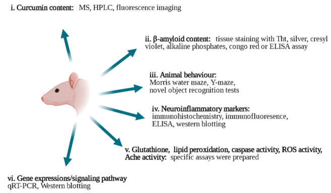
Figure 1.
In vivo characterization techniques applied to study the effects of curcumin on AD (drawn in Biorender). i. Curcumin content: MS [6], HPLC [58,59,75], and fluorescence imaging [21,41] techniques. ii. β-amyloid content: thioflavin T (Tht) staining [61,76,77,78,79] silver staining [39,80], cresyl violet [81], alkaline phosphates [82,83], congo red [78,79], and ELISA assay [2,18,84]. iii. Animal behavior tests: Morris water maze [32,39,85], Y-maze [5,86,87], and novel object recognition tests [5,66]. iv. Neuroinflammatory markers: immunohistochemistry [87,88,89,90], Western blotting [91], qRT-PCR analysis [91,92] and ELISA assay [86,92]. v. Glutathione [3,93], lipid peroxidation [56,87], caspase activity [94,95], ROS activity [96], Ache activity [97,98,99] assays were used. vi. Gene expressions and signaling pathways: qRT-PCR [84,100] and Western blotting [39,84,101].

