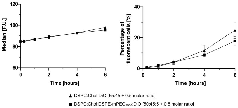
Figure 6.
Cellular uptake of DiO labelled liposomes by 2D LS monocultures. 2D LS monolayers were incubated with non-PEGylated and PEGylated DiO labelled liposomes for 15 min, 1, 2, 4, and 6 h. Cellular uptake was evaluated via flow cytometry. The percentage of fluorescent cells (living gate) showed 24.8 ± 5.2% uptake for non-PEGylated and 17.8 ± 3.0% for PEGylated DiO liposomes. Data are expressed as the mean ± SD (n = 3).

