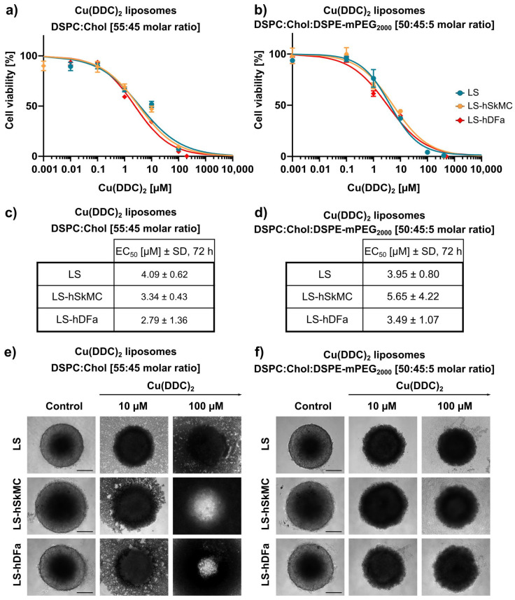
Figure 11.
In vitro cytotoxicity of Cu(DDC)2 liposomes on LS-hSkMC and LS-hDFa co-culture spheroids. (a,b) LS monoculture spheroids, LS-hSkMC and LS-hDFa co-culture spheroids were treated 72 h with non-PEGylated (a) and PEGylated (b) Cu(DDC)2 liposomes. 3D spheroids were generated by using agarose-coated wells with subsequent centrifugation. hSkMC and hDFa cells are mixed with LS cells in a 1:1 ratio for co-culture spheroids, respectively. Cell viability curves were obtained using the 3D CellTiter-Glo® assay after indicated treatment duration. (c,d) EC50 values after 72 h of treatment with non-PEGylated (c) and PEGylated (d) Cu(DDC)2 liposomes. Data are expressed as the mean ± SD (n = 3). (e,f) Representative brightfield microscopy images of LS monoculture and co-culture spheroids with hSkMC and hDFa, respectively. Images were taken after 72 h of treatment with non-PEGylated (e) and PEGylated (f) Cu(DDC)2 liposomes. Scale bar = 200 µm.

