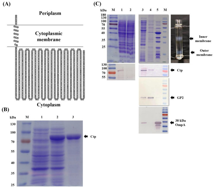
Figure 1.
Ctp protein is localized in the cytoplasmic membrane. (A) A topology model for subcellular localization of Ctp, generated using the SACS MEMSAT server. (B) SDS-PAGE analysis showing M, marker; lanes 1 and 2, uninduced and IPTG-induced E. coli BL21 (DE3), respectively; and lane 3, Ni-NTA His-bind® Resin affinity column purified Ctp protein. Cultures with and without IPTG were normalized to OD600 = 1, and wells were loaded with the same volume of lysate. (C) Top image shows SDS-PAGE analysis: lane 1, crude lysate from ATCC 17978; lane 2, crude lysate from MR14; and lanes 3, 4 and 5, cytoplasm, cytoplasmic membrane and OM proteins, respectively, obtained from ATCC 17978. Image on right shows separation of inner and outer membranes into distinct bands using sucrose gradient. Bottom image: Western blot analysis; each lane corresponds to the respective lane from the top image. Primary antibody of Ctp was used for the blotting assay. Antibodies specific to GP2 and 38 kDa OmpA were used as an internal loading control for inner and outer membrane fractions, respectively.

