Figure 2.
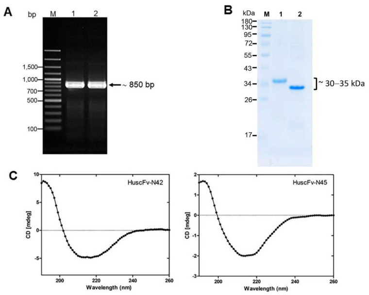
Production of recombinant LasB-bound HuscFvs. (A) Amplicons of huscfv-LIC fragments for sub-cloning into pLATE52 vector. M, 100-bp plus DNA marker; 1 and 2, huscfv-LIC amplicons of the transformed NiCo21(DE3) E. coli clones N42 and N45, respectively. Numbers at the left are DNA sizes in bp. (B) SDS-PAGE analysis of LasB-bound HuscFvs. M, protein marker; 1 and 2, purified HuscFv-N42 and HuscFv-N45, respectively (~30, 35 kDa). Numbers at the left are protein molecular masses in kDa. (C) CD spectra of the refolded HuscFv-N42 (left) and HuscFv-N45 (right).

