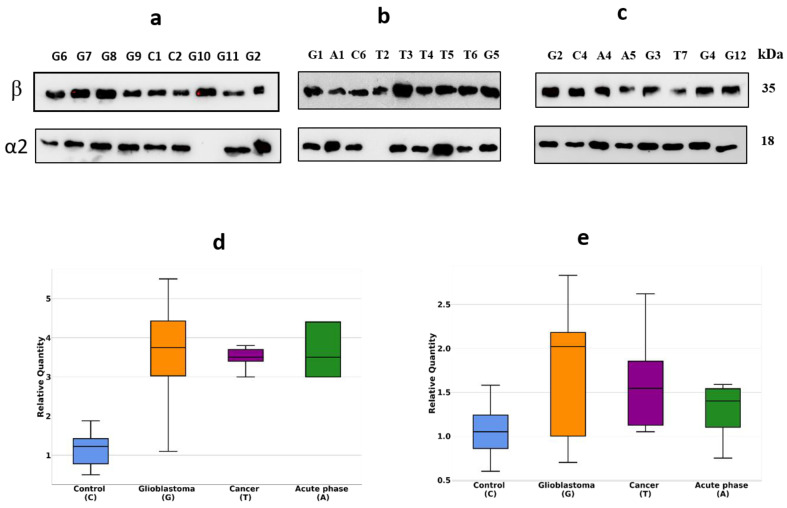
Figure 2.
Western blotting of plasma samples. The duplicates of the gels shown in Figure 1 were tested using Ab against α2-chain and β-chain (a–c). Intensity statistical analysis of the bands of α2-chain and β-chain (d,e). GBM samples are marked as G, control—as C, cancer—as T, acute phase—as A.

