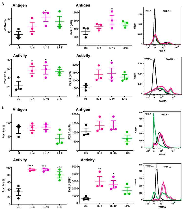
Figure 1.
Active FXIII-A is exposed on the surface of isolated human monocytes and THP-1 cells. (A) Isolated human monocytes or (B) THP-1 cells were left unstimulated or stimulated with IL-4 (20 ng/mL), IL-10 (20 ng/mL) or LPS (100 ng/mL) for 24 h prior to staining using FITC labelled anti-FXIII-A antibody or TAMRA donor FXIII-A substrate for detection of functional activity. Samples were then analyzed using an LSR II flow cytometer. Data are expressed as a percentage of FXIII-A positive monocytes and median fluorescence intensity (MFI) values as the mean ± SEM, n = 3. * p < 0.05; ** p < 0.01; *** p < 0.001 vs. resting cells.

