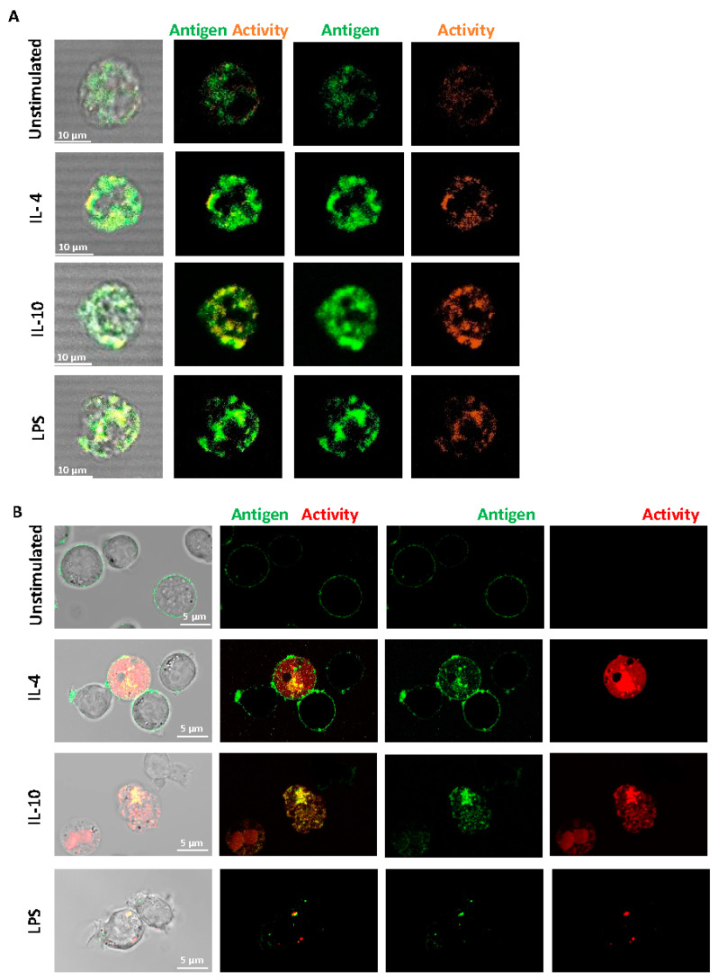
Figure 2.
Distribution of FXIII-A on the surface of isolated human monocytes or THP-1 cells. (A) Isolated human monocytes or (B) THP-1 cells were left unstimulated or stimulated with IL-4 (20 ng/mL), IL-10 (20 ng/mL) or LPS (100 ng/mL) for 24 h. Live cells were stained using FITC labelled anti-FXIII-A antibody or TAMRA donor FXIII-A substrate for detection of activity. Cells were imaged using a LSM880 confocal microscope using a 63 × 1.40 oil immersion. Images are representative of n ≥ 3; scale bar 10 μm (A) or 5 μm (B).

