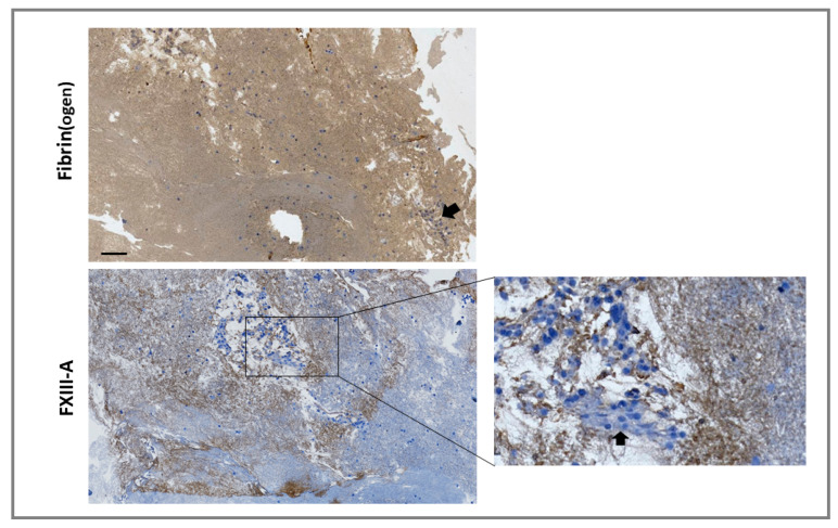
Figure 4.
Monocytes are incorporated into forming thrombi. Model thrombi were formed under arterial shear rates with FXIII–/– plasma in the presence of THP-1 cells. Thrombi were fixed in 10% formalin and wax embedded prior to sectioning. Sections were deparaffinized and stained with a polyclonal rabbit anti-fibrin(ogen) (top panel) or anti-FXIII-A (bottom panel) antibodies (brown) and counterstained with Mayer’s haematoxylin (blue) for detection of the nuclei of THP-1 cells. Imaged using 5× magnification. Cell clusters were detected in certain locations (inset and as indicated by arrows). Scale bar = 100 µm.

