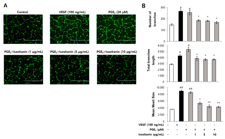
Figure 3.
The effect of isookanin on tube formation in PGE2-induced HMEC-1 cells. Cells were pretreated with the indicated concentrations of isookanin for 2 h before stimulation with PGE2 (20 µM) or VEGF (100 ng/mL) for 24 h. HMEC-1 cells tube formation was measured using the tube formation assay. (A) Calcein AM dye-stained cells were photographed under a microscope; scale bars are 1000 μm. (B) Tube formation was evaluated by analyzing the total branch number, length, and mesh size of the tube-like structures using Image-J software. The results are mean ± standard deviation (SD) (n = 3). # p < 0.05, ## p < 0.01 vs. control. * p < 0.05, ** p < 0.01 vs. PGE2-treated control.

