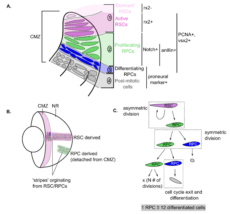
Figure 2.
Retinal stem and progenitor cells’ characteristics in the CMZ of teleost fish and Xenopus. (A) Schematic of the four main compartments of the CMZ niche. The first region at the tip (1) contains dormant and active retinal stem cells (RSCs) (pink) that are either rx2- or rx2+. In the second region are highly proliferative retinal progenitor cells (RPCs) that are distinguished by expression of a different subset of markers (i.e., Notch and Anillin). The third region is composed of RPCs poised to differentiate since they co-express proliferating and pro-neural genes. All actively proliferating RSC/RPCs express markers for proliferation, including PCNA and vsx2. In the fourth region are post-mitotic cells that have differentiated. This schematic is adapted from those seen in [6,7]. (B) Schematic of the whole eye showing the CMZ region labelled in pink in an annulus adjacent to the lens. Labelled populations of cells derived from RSCs produce continuous stripes from the CMZ (named ArCoSs), whereas stripes produced from RPCs produce disconnected patches called terminated patches. This schematic is adapted from data visualized in [41,46,48]. (C) Example of the different events occurring in the proliferation and differentiation of RSCs/RPCs in the CMZ. An asymmetric division gives rise to two different daughter cells. An RSC that asymmetrically divides gives rise to one RPC and one RSC to maintain self-renewal. Conversely, a symmetric division results in the progeny of the same identity (i.e., two RPCs). RPCs will eventually stop proliferating and differentiate after a certain number of divisions, on average producing 12 differentiated cells. Pink cells = RSCs (zone 1 cells), green cells = proliferating RPCs (zone 2 cells), blue cells = RPCs committed to differentiation (zone 3 cells) and grey cells = differentiated cells. Adapted from data presented in [42].

