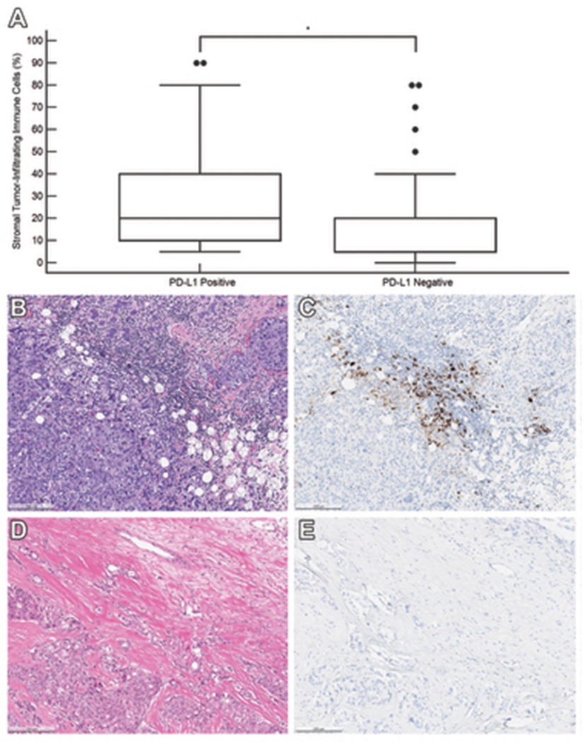
Figure 1.

Association between stromal tumor-infiltrating immune cells and PD-L1 (SP142) immunohistochemistry result in 156 triple-negative breast carcinoma samples. (A) Box and whisker plots of the percentage of stromal area occupied by immune cells demonstrates a significantly higher percentage in PD-L1 positive tumors than in PD-L1 negative tumors (P < .001). * denotes significant P < .05. (B) Case of a 32-year-old woman with primary invasive carcinoma of no special type, harboring stromal-infiltrating immune cells that occupy 60% of the stroma and (C) express PD-L1 [PD-L1 (SP142) immunohistochemical stain]. (D) Case of a 55-year-old woman with primary invasive carcinoma of no special type, status-post neoadjuvant chemotherapy. The stromal area is densely collagenous with 5% of the stroma occupied by immune cells with (E) no expression of PD-L1 [PD-L1 (SP142) immunohistochemical stain].
