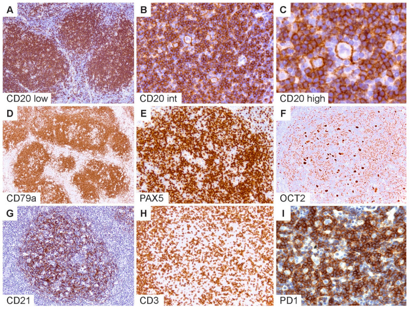
Figure 2.
Immunophenotypic features of NLPHL. (A) CD20 highlights numerous classic lymphoid nodules. (B,C) Scattered LP cells reside within nodules containing a microenvironment rich in small B-lymphocytes. (D) CD79a, (E) PAX5 and (F) OCT2 show staining of LP cells and the surrounding small B cells. (G) CD21 defines an intact follicular dendritic cell meshwork within an NLPHL nodule. (H) CD3 stains background T cells with occasional ring formations and (I) prominent PD1-positive rings surrounding LP cells. [Original magnifications: A,D,G,H ×60; B,E,F,I ×150; C ×600].

