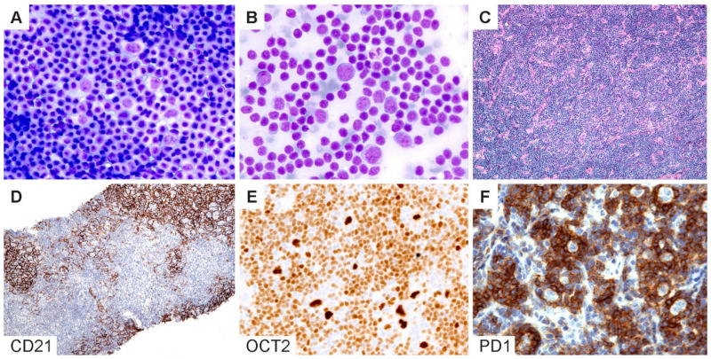
Figure 6.
Cytologic diagnosis of NLPHL. (A,B) Wright–Giemsa-stained touch preparations show LP cells in a background of small lymphoid cells and occasional inflammatory cells. (C) H&E section of a core needle biopsy does not readily reveal a nodular architecture. (D) A CD21 stain highlights the FDC meshworks and confirms the presence of nodules. (E) Bright OCT2 staining and (F) prominent PD1-positive rings lend support for the diagnosis of NLPHL. [Original magnifications: A,B ×600; C,D ×40; E,F ×150].

