Figure 3.
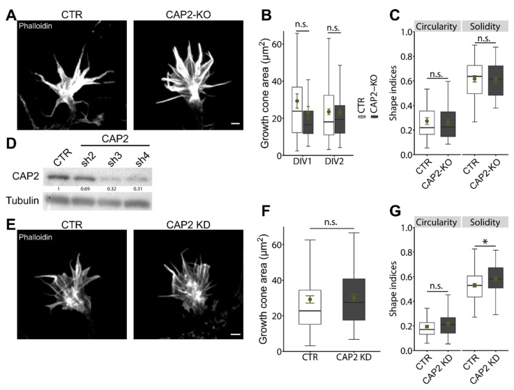
Normal growth cone size and morphology in CAP2-KO neurons. (A) Micrographs of phalloidin-stained growth cones from stage 2 CTR and CAP2-KO neurons. (B) Growth cone size in stage 2 CTR and CAP2-KO neurons at DIV1 and DIV2. (C) Growth cone shape indices for circularity and solidity for stage 2 CTR and CAP2-KO neurons. (D) Immunoblots with lysates from cerebral cortex neurons nucleofected with three different shRNAs directed against CAP2 (CAP2-sh2, CAP2-sh3, and CAP2-sh4) or with a control (CTR) shRNA. Tubulin was used as the loading control. Values indicate CAP2 levels in CAP2-KD neurons relative to CTR-shRNA-nucleofected controls. (E) Micrographs of phalloidin-stained growth cones from replated stage 2 neurons nucleofected with either CTR shRNA or a mixture of CAP2-sh3 and CAP2-sh4 (CAP2-KD). (F) Growth cone size and (G) shape indices for circularity and solidity in stage 2 neurons nucleofected with either CTR shRNA or CAP2-sh3/CAP2-sh4. Scale bars (in µm): 2 (A), 2 (E). * p < 0.05, n.s.: p ≥ 0.05. Error bars represent SEM.

