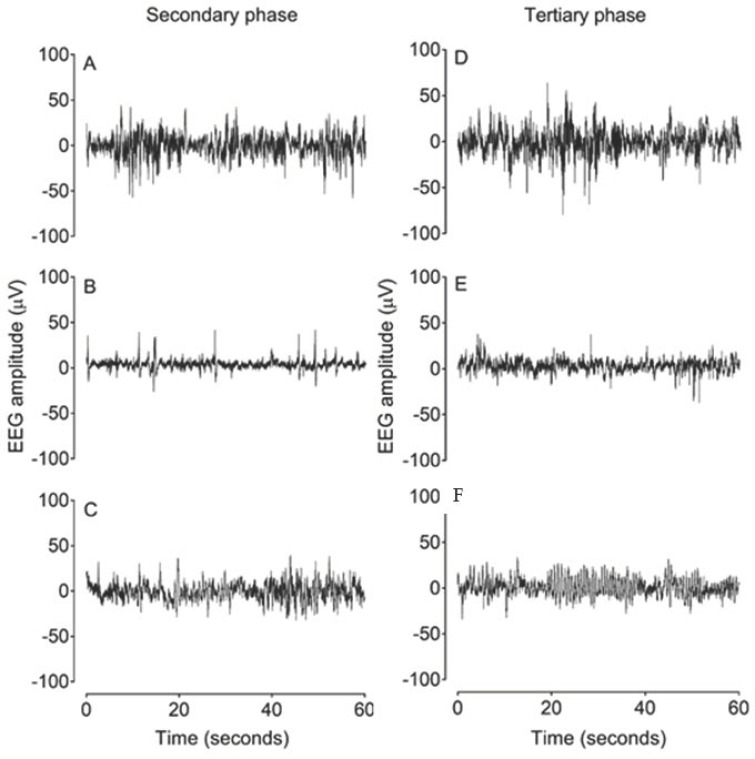
Figure 3.
Examples of raw EEG activity from sham asphyxia, asphyxia + vehicle and asphyxia + GDQ groups in the secondary phase at 18 h after umbilical cord occlusion (UCO) (A–C) and in the tertiary phase at 84 h after UCO (D–F). Panels (A) and (D) demonstrate normal discontinuous mixed amplitude and frequency observed in preterm fetal sheep at 0.7 gestation. Panel (B) demonstrates suppressed background activity interspersed with the presence of sharp and fast wave transients in the asphyxia + vehicle group in the secondary phase. Panel (C) shows greater background activity of higher amplitude in the asphyxia + GDQ group. Panel (E) demonstrates a mildly suppressed background activity in the asphyxia + vehicle group in the tertiary phase. Panel (C) shows that the reduction in SEF seen in the asphyxia + GDQ group in Figure 2 is associated with slow rhythmic epileptiform discharge (ED) activity.

