Figure 1.
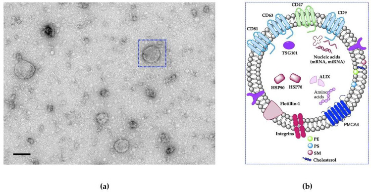
Natural EVs morphology. (a) TEM image of natural EVs (oviductosome, OVS) isolated from mouse oviductal fluid of ~100 nm in size. Scale bar represents 100 nm. (b) Schematic showing the structural components and cargo of extracellular vesicles which packed with a variety of cellular components, including various transmembrane proteins (tetraspanins, PMCA4), heat shock proteins, adhesion proteins, nucleic acids (mRNAs and miRNAs), and lipids (PE, PS, SM, and cholesterol). EVs also protect encapsulated cargo from clearance by macrophages via CD47 (“do not eat me” signal).

