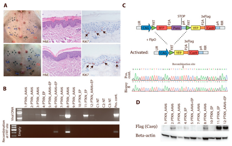
Figure 4.
In vivo activation of Cas9 expression. Cas9 minipigs were transduced in multiple areas in the abdominal skin, and biopsies were taken after 1 month. (A) Tissue sections from transduced areas and control skin were stained for H&E and Ki67 (n > 3). (B) Biopsies were analysed for the presence of viral DNA and for recombination at the FRT sites and hereby activation of Cas9 expression. The PTKN construct was used as either plasmid or in AAV6 particles. Some areas underwent electroporation (EP) to enhance the delivery. NT: nontreated. (C) Two samples underwent Sanger sequencing to confirm recombination of the transposon at the FRT sites. (D) Proteins isolated from the biopsies were analysed by Western blotting for Cas9-Flag expression. Beta-actin was used as loading control. Western Blot Images can be found in Figure S7.

