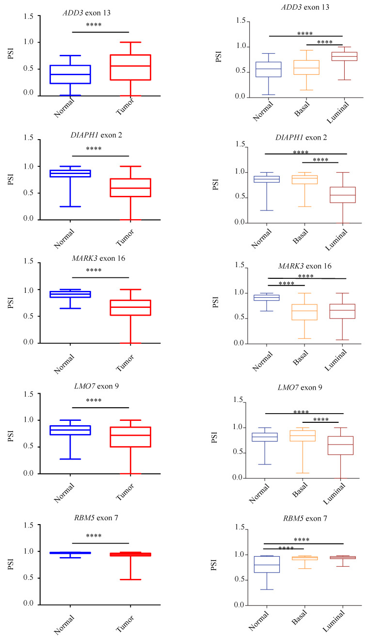
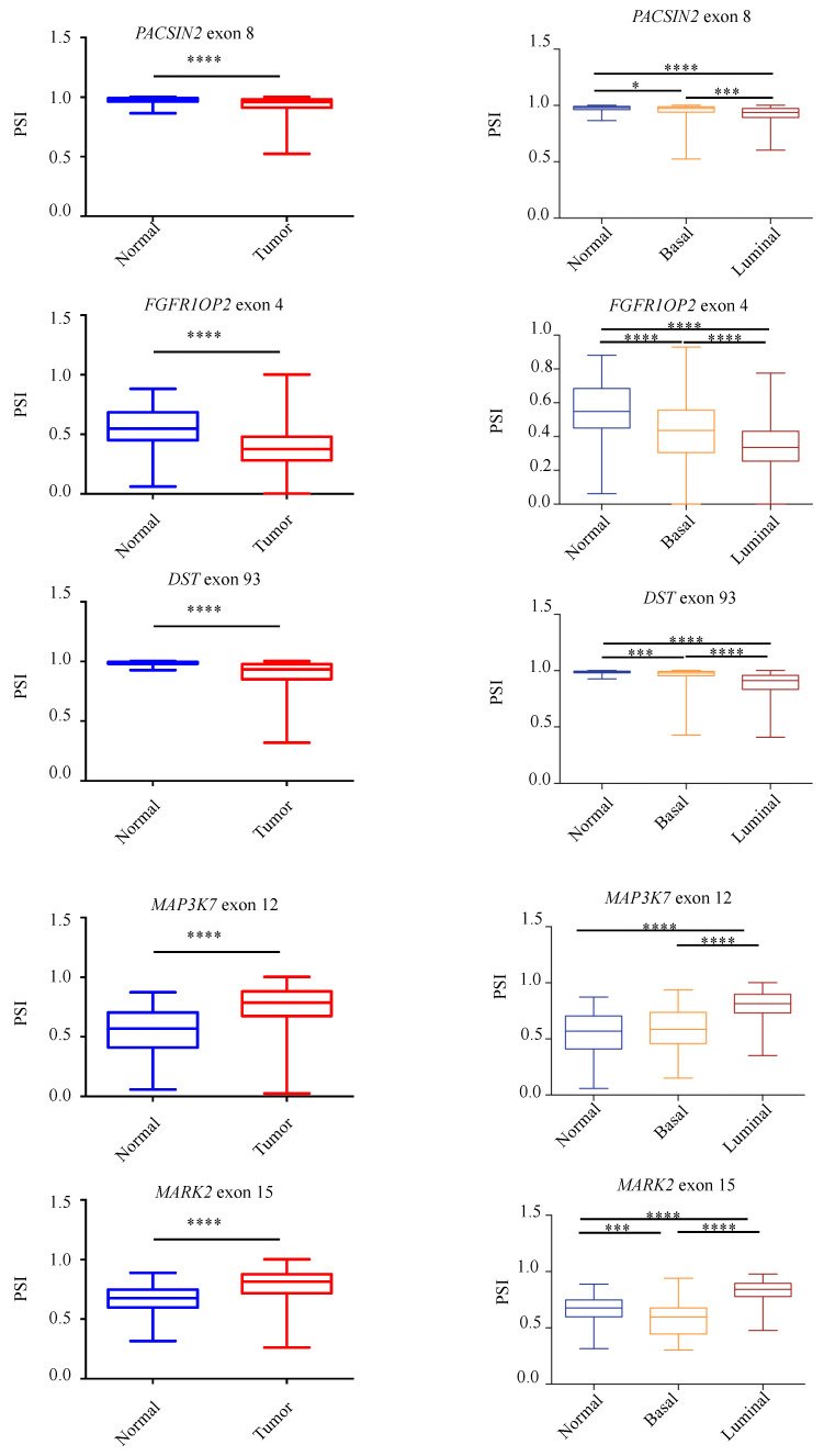
Figure 5.
Validation of RASL-seq AS events in breast cancer specimens and in non-pathological tissues. Analysis, by using the TCGA SpliceSeq online tool, of the splicing pattern of ADD3 exon 13, PACSIN2 exon 8, DIAPH1 exon 2, FGFR1OP2 exon 4, MARK3 exon 16, DST exon 93, LMO7 exon 9, MAP3K7 exon 12, RBM5 exon 7, and MARK2 exon 15 in normal and primary tumor specimens from the TCGA-BRCA dataset (left) and in normal and primary tumor specimens divided for their histopathological state (basal or luminal) (right). PSI = Percent Splice-In. **** p < 0.0001, *** p < 0.001, * p < 0.05.


