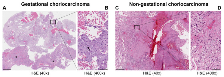
Figure 1.
Representative images of hematoxylin and eosin (H&E) staining in gestational (A,B) and non-gestational (C,D) choriocarcinoma. Uterine curettage (A,B); (A) low power (40×; scale bar = 500 μm) with abundant decidualized endometrium and blood and two large clusters of choriocarcinoma (*); (B) high power (400×; scale bar = 50 μm) shows aggregates of large trophoblasts with marked atypia and prominent mitotic figures (arrow) covered by atypical syncytiotrophoblasts. Invasive choriocarcinoma in the ovary (C,D). (C) Component of a germ cell tumor of the ovary with large and atypical trophloblastic cells with hemorrhage and necrosis (low power 40×; scale bar = 500 μm) associated with dysgerminoma (not present in the picture). (D) Markedly atypical synciotrophoblasts and cytotrophoblasts (high power 400×; scale bar = 50 μm).

