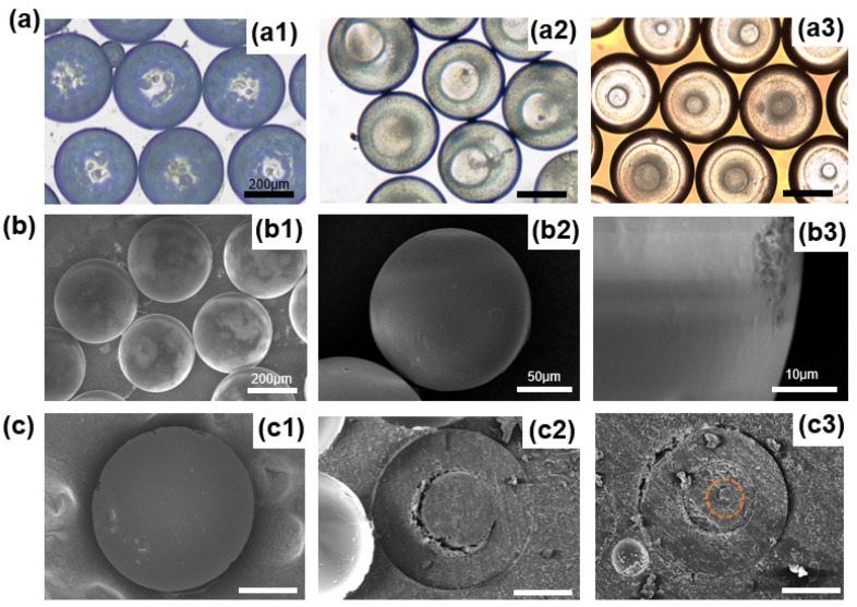
Figure 5.
(a) Optical microscopic images of microspheres with different numbers of layers: (a1) One layer, (a2) Two layers, (a3) Three layers. The scale bar is 200 µm. (b) Scanning electron microscope images of microspheres prepared at one flow rate ratio. (b1) SEM image showing good monodispersity and sphericity of microspheres; (b2) Magnified SEM image of the microsphere; (b3) SEM image showing the solid, smooth, and dense surface of the microsphere. (c) Scanning electron microscope images of different number-layered microspheres cut open: (c1) One layer, (c2) Two layers, (c3) Three layers. The scale bar is 100 µm.

