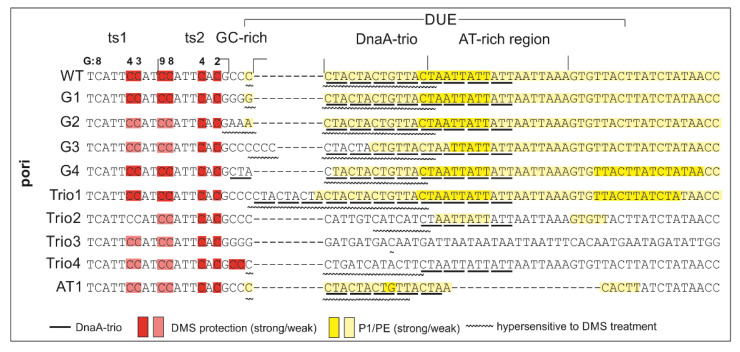
Figure 4.
Summary of DnaA-trio analyses in H. pylori conducted in this study. The most important features of the analysis are indicated as follows: the red and pink rectangles depict protection of DnaA boxes upon protein binding and decreased interaction in comparison with the poriWT sequence, respectively; the intensity of the yellow rectangles indicates the susceptibility of DNA strands to P1 nuclease digestion and the frequency of the open complex formation; wavy lines indicate sequences that were hypersensitive to DMS methylation upon DnaA binding.

