Figure 9.
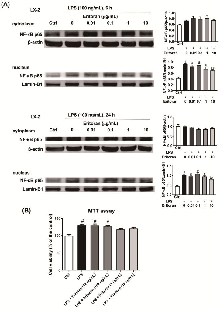
The NF-κB p65 nuclear translocation in LX-2 cells. (A) Incubation of LX-2 cells (2 × 10⁶/well) with lipopolysaccharide (LPS, 100 ng/mL) and with a serial dose of eritoran (0–10 μg/mL) for 6 h and 24 h. Western blotting of nuclear and cytoplasmic protein of NF-κB p65 was performed (n = 4/group). (B) Cell viability determined by the methyl thiazolyl tetrazolium (MTT) assay in LX-2 cells (1 × 10⁴/well) incubated with the control medium (Ctrl), LPS (100 ng/mL) and a serial concentration of eritoran (0–10 μg/mL) for 24 h (n = 8/group); # p < 0.05 vs. the Ctrl group; * p < 0.05 vs. the group treated with LPS and without eritoran.

