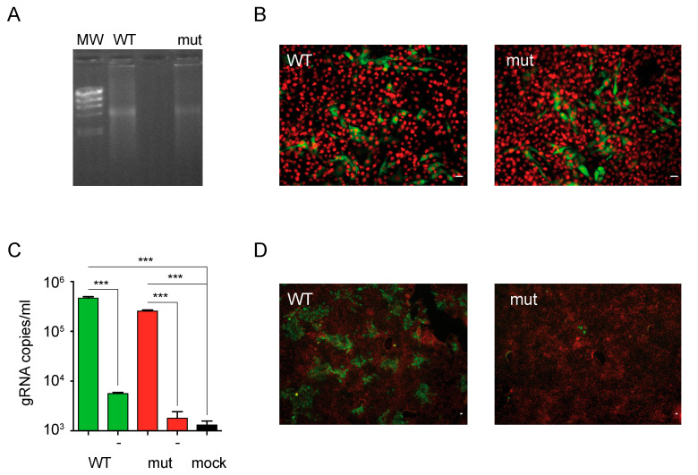
Figure 4.
BVDV particles bearing mutations in the exposed β-hairpin motif display reduced infectivity. (A) Agarose gel electrophoresis of in vitro transcribed genomic RNAs of wild type (WT) and mutant (mut) ncpBVDV. Lambda DNA/HindIII marker (MW) was used as a reference. (B) E2 expression in cells transfected with wild type and mutant RNAs. Representative fluorescence microscopy images acquired with 20× objective of MDBK cells transfected with wild type (WT) or mutant (mut) RNAs and stained with a polyclonal antibody against E2 (green channel) and DAPI (red channel). Scale bar: 10 µm. (C) BVDV virion production to the supernatant of transfected cells. Bar graph for the titers of wild type and mutant viruses produced to the supernatant of transfected cells estimated by qPCR. No reverse transcriptase controls are indicated with (-). The black bar represents a control for mock transfected cells. Error bars represent the standard deviation of triplicate points. Data were analyzed by one-way ANOVA with Bonferroni’s posttest (***, p < 0.001). (D) Infectivity of wild type and mutant BVDV virions. Representative fluorescence microscopy images acquired with 4× objective of MDBK cells infected with wild type (WT) or mutant (mut) BVDVs recovered from transfected cells and stained with a polyclonal antibody against E2 (green channel) and DAPI (red channel). Scale bar: 10 µm. The results are representative of at least three independent transfections.

