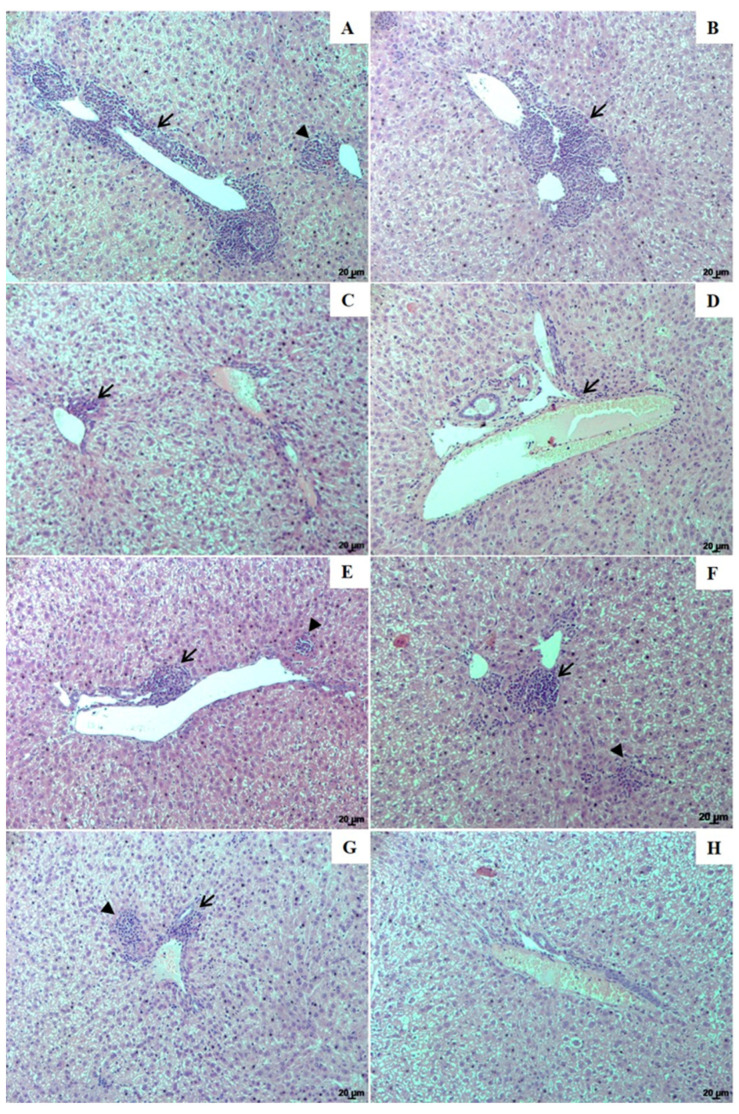
Figure 6.
Histological changes of the liver from golden hamsters infected with L. (L.) infantum. Liver histological sections from (A)—infected control; (B)—infected and treated with empty NLC; animals treated with 1.25 and 5.0 mg/kg UA loaded in NLC (C,D, respectively), animals treated with 1.25 and 5.0 mg/kg free UA (E,F, respectively) or AmB (G). Liver histological sections from healthy animals are shown in image H. Inflammation foci (arrows) and granulomas (arrowhead). Magnification of 100×; scale bars: 20 μm (A–H).

