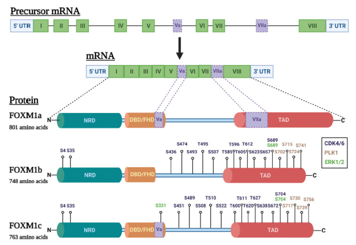
Figure 2.
FOXM1 isoforms and phosphorylation sites in ovarian cancer. Top: FOXM1 precursor mRNA (with introns and exons indicated) followed by FOXM1 mRNA structure (exons only). Exons shared by all FOXM1 isoforms are shown in green while alternative exons are shown in light purple. Bottom: Diagram of protein structure of the three major FOXM1 isoforms: (1) FOXM1a, which contains alternative exons Va and VIIa; (2) FOXM1b, which contains no alternative exons; and (3) FOXM1c, which contains alternative exon Va. The three major protein domains are indicated: N-terminal repressor domain (NRD, teal); DNA binding/forkhead domain (DBD/FHD, orange); and transactivation domain (TAD, red). The protein regions corresponding to the alternative exons Va and VIIa are shown in light purple. FOXM1 residues reported to be phosphorylated by three kinases important in ovarian cancer, CDK4/6 (dark blue), PLK1 (tan), and ERK1/2 (green), are indicated. Figure created with BioRender.com.

