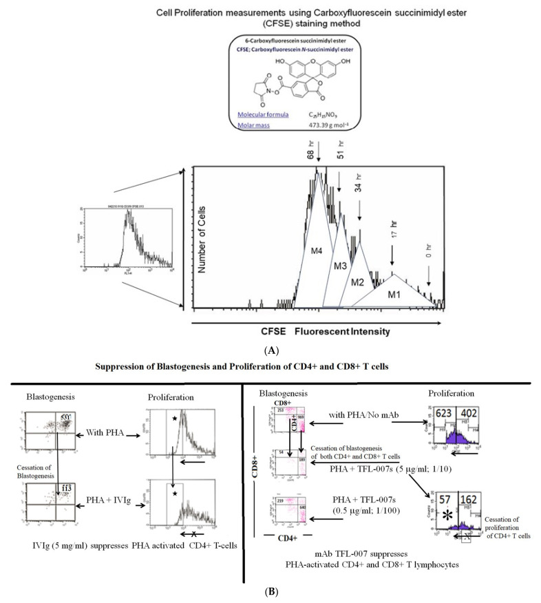
Figure 6.
(A). The proliferation assay is based on labeling the purified T-cells during PHA activation with the intracellular fluorescent dye carboxyfluorescein succinimidyl ester (CFSE: C25H15NO9; mol. mass: 473.39 g/mol) and using flow cytometry, measuring mitotic activity by the successive twofold reductions in fluorescent intensity of the T-cells placed in culture for 72 h. CFSE is cell-permeable and is retained for long periods within cells by covalently coupling by means of its succinimidyl group to intracellular molecules. Due to this stable linkage, once incorporated within cells, CFSE is not transferred to adjacent T-cells but remains in the cell even after several mitotic divisions. (B). Suppression of blastogenesis and proliferation of CD4+ T-cells by IVIg (Globex) and HLA-I polyreactive mAb TFL-007 at similar protein concentrations. The CFSC profile illustrates suppression as indicated by asterisks.

