Figure 3.
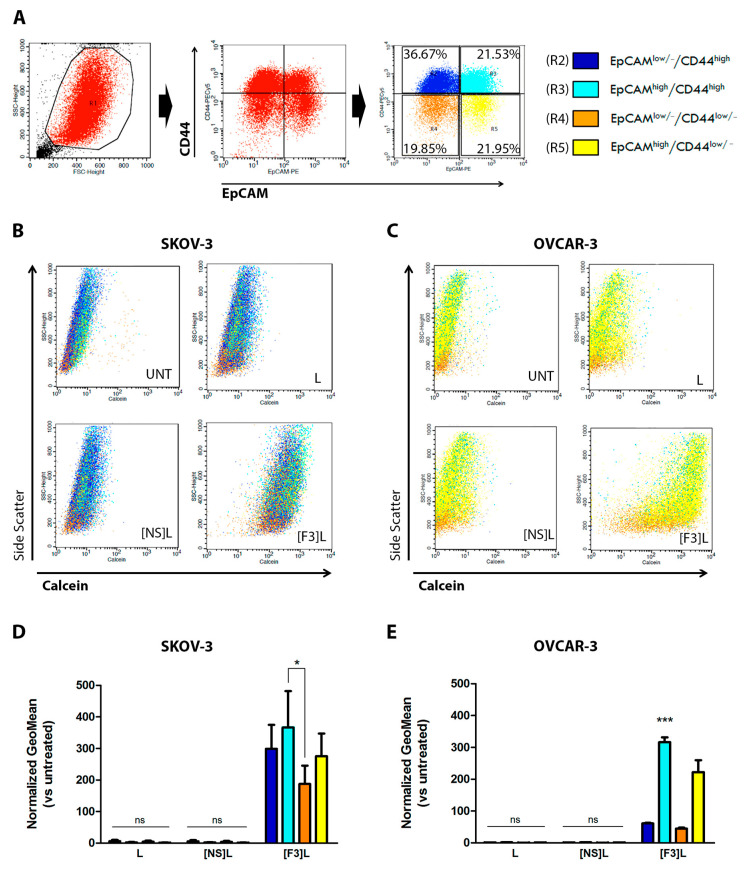
Cellular association of F3 peptide-targeted liposomes with putative ovarian cancer stem cells. (A) Flow cytometry gating strategy for the identification of CSC-enriched (R3, light blue) and non-stem cancer cells, -SCC (R4, orange) sub-populations based on the assessment of CD44 and EpCAM surface levels is represented. The calcein/side scatter dot-plots reflecting the calcein signal distribution for (B) SKOV-3 and (C) OVCAR-3 bulk cells is represented, upon incubation with 0.4 mM (total lipid) of calcein-labeled F3 peptide-targeted ([F3]L), non-specific peptide targeted ([NS]L) or non-targeted (L) liposomes for 4 h at 37 °C. The geometric mean of calcein fluorescence levels for each of the indicated sub-populations for (D) SKOV-3 and (E) OVCAR-3 cell lines are represented. Data represent the mean ± SEM (n = 3; two-way ANOVA with Bonferroni’s post-test; ns p > 0.05; * p < 0.05 and *** p < 0.001).

